Samir Dahouk
Seaside, CA
![]()
Samir Dahouk
Seaside, CA
###The following information is targeted toward families of SMA1, their friends, and
doctors alike. Contents are derived from Samir's medical records and the parents'
experience. We hope you find it informative and helpful.###
***This Page is Under Construction***
Let me know what you think about my page. Send mail by clicking here.
Preface: We have lost the battle against Spinal Muscular Atrophy
(SMA I) on Thursday 12 November 1998. After 7 1/2 months of constant battle at home and in
9 different hospitals in Germany and the US, Samir, our son, was no longer capable of
fighting. He passed away at home peacefully between the arms of his mother.
In the past 2 months, after Samir's last hospitalization at Travis AFB, CA and then
settling at DLI in Monterey, Mary and I worked very hard to keep him with us. Our home
became a 24 hours mini intensive care unit - Oxygen tanks (to supplement his shallow
breathing), heart and oxygen blood level monitors, Bi-PAP machine (to help him breath when
he's tired), suction machine ( to suction his mouth and throat area), inhalation machine
for chest therapy, kangaroo pump ( to pump milk into his stomach thru a nasogastric tube),
Ambu bag (for mouth resuscitation when he struggles to breath), chest physical therapy (to
move any fluids/mucus lodged in his chest) and many medicines to include Morphine (for air
hunger pain). But the Will of God is greater than all and It must prevail. God took him
away young and innocent to escape the envy of man and the evils and sorrows of this
present world.
On 9 November 1998, we accepted Hospice care which increased the nursing hours to 8 per
day. But, by accepting Hospice, we were denied any heroic measures to save Samir's life to
include CPR or dialing 911. Hospice is for the terminally ill and dialing 911 is a breach
of contract !! Anyway, Samir gave us all the signs that he's no longer capable of
sustaining life. Besides his progressive muscle weakness due to SMA, Morphine was not
enough to ease his pain/air hunger; his CO2 level in his blood reached a poisoning level;
his body began to swell...
What is SMA I?
Spinal muscular atrophy is the leading genetic cause of infant death. This neuromuscular
disease, characterized by the degeneration of motor nerve cells that control the body's
voluntary muscles from the head down, originates from defects in the Survival of Motor
Neuron (SMN) gene on chromosome 5. Spinal muscular atrophy is an inherited condition that
affects about one in 6,000.
How was he diagnoed with Type I Spinal Muscular Atrophy - Werdnig-Hofmann?
During a routine screening, Samir was found to present with marked hypotonia and
hardly elicitable reflexes. So, he was admitted at Friedrich-Alexander Univercity
Children's Hospital in Erlangen Germany and received inpatient care from 19 June to 1 July
1998. He is the third child of a healthy couple. Spontaneous delivary took place at 39
weeks gestation. Birth wt 3540g; Ht 54 cms. He has a 3 1/2 year old sister who is well. A
brother who was a preemie grower of 24 weeks gestation died at age of 4 weeks due to
marked bronchopulmonary dysplasia and bilateral intraventricular hemorrhage with the
possibility of SMA1.
Condition on Admission: 11-week-old male infant; tachydyspnea at a respiratory
rate of 80/min; tongue fasciculation; poor muscle tone; proprioceptive reflexes could not
be elicited; however, the infant was awake and alert, seeking eye contact, fixing and
laughing. Liver palpable 4 cm and spleen 1 cm below thw costal margin; diminished
spontaneous motor activity. Skin unremarkable. Normal pupillo-ocular motor activity; no
enlarged lymph nodes; cardiac action clean and regular; lungs symmetrically ventilated;
clear breath sound. Male infantile genitalia; oropharyngeal region irritation-free. Weight
on admission 5160 g.
Laboratory Tests: Hemoglobin 11.4 g/dl; erythrocytes 4.14 million/ul, leukocytes
7.100/ul; hematrocrit 34.9%; platelets 272,000/ul. Normal differential blood count and
blood gases analysis based on Astrup. Blood suger, ionogram, retention parameters, and
phosphate also within the limits of normal. Aspartate aminotransferase 14 U/I; alanine
aminotranferase 14 U/l; gamma glutamyltransferase 12 U/l; alkaline phosphatase 343 U/l;
creatine kinase 56 U/l; C-reactive protein 0.2 ng/dl; Quick (one-stage prothrombin time
test) 97%;partial thromboplastin time 42.1%. Serum phenylalenine concentration 0.4 mg/dl;
urine analysis NAD.
Immunoglobulins: lgA 37 mg/dl; lgG 382 mg/dl; lgM 33 mg/dl. Transferrin 21 mg/dl.
Triglycerides 124 mg/dl; cholesterol 115 mg/dl; hdl 31.8 mg/dl.
Lumbar Puncture: 1 leukocyte; total protein in the CSF 33 mg/dl; CSF lactate 0.82
mg/dl.
Thyroid parameters: fT4 21.6 pg/ml; TSH 3.6 uU/ml (euthroidism).
Diagnostic investigation of metabolic status: Serum ammonia concentration 74.8
mg/dl; serum lactate 0.8 mg/dl; mucopolysaccharides in the urine NAD; origanic acids in
the urine NAD. Overlong chain fatty acids in the plasma: all values were within the limits
of normal. Leukocyte preparation: no signs of metachromatic leukodystrophy, GM1
gangliosidosis, GM2 gangliosidosis variant B or Sandhoff disease (GM2 gangliosidosis
variant 0)
Cardio investigation: ECG on 12 June 98: Sinus rhythm; verical type; signs of
still age appropriate biventricular stress; no depolarization distribances; no cardiac
irregularity. Another ECG on 19 June: No vitium cordis but myocardial hypertrophy of the
ventricle; no valvular problems.
Neurological investigation: EEG on 22 Jun 98: Age-appropriate electroencephalogram
which was partly hampered by movement artifacts; no hypersynchronous activity or focal
sites.
NCS on 19 Jun 98: Pathological NCS with an abnormally high stimulation threshold and
reduced amplitude and nerve conduction velocity.
EMG: Abnormal for polyphasia and insertional activity; no spontaneous activity.
EMG on 26 Jun 98: Pathological EMG; right medial rectus muscle with sparse spontaneous
motor activity and a definitly pathological neurogenic high- polyphasic pattern associated
with voluntary motor activity.
AEP on 21 Jun 98: prolonged latency on both sides; no signs of a hearing loss.
SEP on 3 Jul 98: Pathological SEP with a prolonged nuchal tendency, diminished sensory
nerve conduction velocity and only coarse cortical negativations on the right; no cortical
potential on the left. Suggests involvement of central myelinization.
Ophthalmological consultation
on 21 Jun 98: OD/OS: Pink optic disk, sharply defined, level, macula NAD.
Ultrasound investigation:
Hips on 19 Jun 98: type lb hips on both sides based on graf. Abdomen on 19 Jun 98: Liver 2
cm below costal margin, normal structure; spleen NAD; kidneys NAD. Small pericardial rim,
non-circular, max. 3.9 ml. CNS on 22 jun 98: CNS structurally NAD.
Radiologic investigations:
Skeletal X-ray of the skull, spinal column, pelvis and right wrist: Based on the skeletal
X-ray taken there were no signs of skeletal dysplasia.
Chest X-ray on 19 Jun 98: Bell-shaped chest; broad lower thoracic aperture; skin fold
overlapping toward the right lateral aspect; well defined enlargement of the middle shadow
to the left. Thymus enlarged. Hilus a bit accentuated and ill-defined on both sides with
slightly increased bronchitic and peribronchitic marking of the medial lower field
sections. Comparatively flat diaphragm on both sides.
Histologic report on muscular biopsy on 28 Jun 98
Sent was muscle tissue in formalin, muscle we found large fields of atrophic fibres. Their
diameters in the paraffin sections with HE stain were ranging between 7 and 20 um. No
clear increase in nuclei.
These atrophic fields contained small groups of normal-calibered and hypertrophic fibers
whic were 40-60 um in diameter. No structural changes demonstrable in the fibers. The
connective tissue in the interstitium was smooth; no excessive tissue. No lipomatous
transformation. Normal muscular vessels. No inflammstory changes.
Conclusion: The extreme difference in size between the fields of atrophic fibers and
groups of normal-calibered and hypertrophic fibers points to a neurogenic muscular
affection. The enlarged, hypertrophic fibersare suggestive of congital mascular dystrophy.
However, structural changes were absent in the fibers. The absence of connective or
lipomatous transformation is an indication that there is no such medical condition. On the
whole, this is the most likely a case of spinal infantile muscular atrophy. If this
suspicioncannot be confirmed by genetic study, hereditary neuropathy should be considered.
A final diagnosis cannot be made as yet. the report of the nerve biopsy will be in on 17
jul 98. We will contact you immediately afterwards.
Muscle bioppsy specimen with advanced field atrophy, most likely a case of spinal
infantile muscular atrophy.
Molecular genetic diagnostic investigation on 8 Jul 98: Evidence of homozygous
deletion of exons 7 and 8 in the telomeric copy of the SMN (survival motoneuron gene) in
this infant which confirmed the tentative diagnosis of infantile spinal muscular atrophy
from a molecular genetic viewpoint.
Treatment and progress: Based on the clinical signs including marked muscular
hypotonia, distinctive tongue fasciculation, poor feeding effort and faint crying in this
nonetheless very alert infant who also was responding well when talked to a condition of a
spinal muscular atrophy was strongly suspected which was then confirmed by a muscular
biopsy and genetic investigations. Because the first electromyogram was not were
conclusive and in view of the additionally noted mild hepatomegaly and hypertrophic
cardiomyopathy we carried out additional diagnostic investigations with regard to the
infant's metabolic status but the tests, on the whole, did not reveal any abnormalities.
The AEP and SEP results may indicate that Samir is not only suffering from spinal muscular
atrophy but also a central myelinization disorder. However, in order to confirm this
additional suspicion, we will first wait for the result of the electron microscope study
of the surel nerve biopsy.
Father's Fairwell Remarks at the Cemetery:
In The Name of God the Merciful the Compassionate
Thanks to the ALMIGHTY - Lord of the Universe
Samir, my beloved son, you have touched the hearts and minds of many different doctors,
nurses and staff of 9 different hospitals, the staff and nurses of VNA and Hospice,
friends, neighbors, and family alike. Despite you pain, you have greeted everyone with a
smile without prejudice.
In the past 2 months, we worked very hard to keep you with us, and yes, you worked harder.
But the Will of God almighty is great and His Will must prevail. Your mom, sister Sada,
and I cannot defy the Will of God. Hence, we must let you go now so you can rest in peace
forever. Your body is torn but your soul and memories will remain evermore among us. So, I
turn to Mary the Mother of Jesus and say our son is gone too and in this earthly life we
shell never see our dear one again. And still, we would not have it otherwise because we
know that God wished to take Sammy young and innocent to escape the envy of man and the
sorrows and evils of this present world. And we are so grateful for the assurance that
Sammy is safe and painless in Heaven. So, God grant us the strength, comfort, and wisdom
that we so greatly need on earth to remain humbly submissive to the end.
Samir, you are too pure, too lovely to live on earth.
Fairwell to you my son. May you rest in peace now.
Your proud dad.
Samir and big sister Sada-6 weeks old |
Samir and Daddy-3 months old |
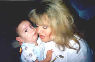
Samir and Mommie-7 months old |
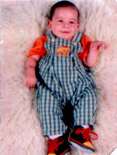
6 1/2 months old |
Families of SMA Home Page
SMA Foundation
This page has been visited
times.

Supported by SMA Support.